December 21, 2020
Hitachi, Ltd., through collaboration with Hiroshima University and Hibino Hospital (Hiroshima, Japan), has developed a technology to estimate the lesion responsible for depression-related symptoms for stroke patients. The right Rolandic operculum*1 was shown to have an association with depression-related symptoms; lesions in the right Rolandic operculum may be related to the pathophysiology of post-stroke depression. The developed technology follows a data-driven*2 approach, which classifies patients in accordance with similarities concerning multiple depression-related indicators, and analyzes lesions using MRI brain images. This technology is expected to assist doctors in providing better care for patients. In the future, Hitachi will implement this technology to investigate not only depression-related symptoms but also a variety of conditions in cerebral infarction patients. By promoting the use of this technology in rehabilitation facilities and hospitals Hitachi aims to improve the quality of life (QoL) of patients.
A technique for estimating the area of the brain associated with depression-related symptoms by analyzing lesions identified from MRI brain images and a data-driven categorization for patients based on similarities concerning multiple depression-related indicators.
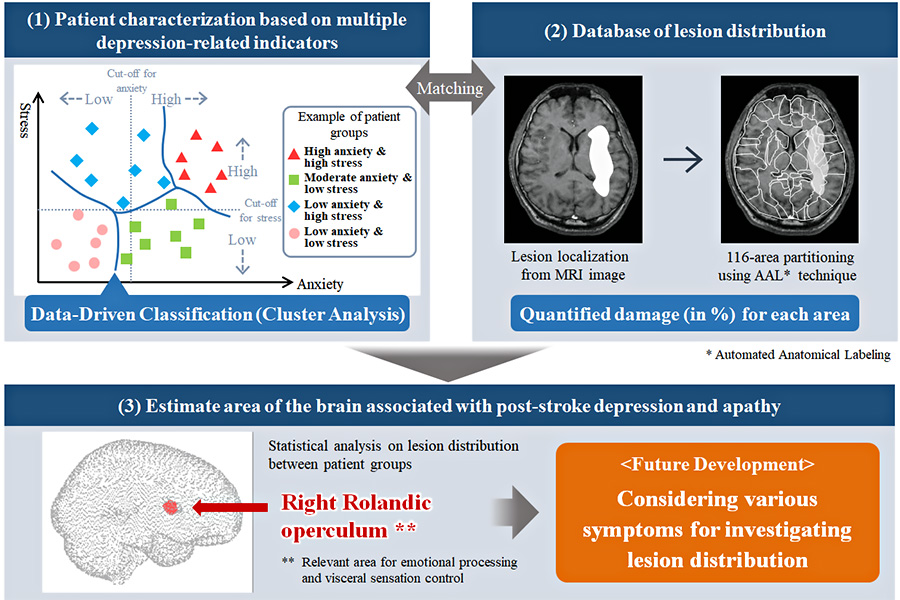
The data-driven analysis of multiple depression-related indicators (e.g., stress, depression, apathy, and anxiety) has shown that cerebral infarction patients can be classified into four groups. Between-group comparisons revealed that lesions in the right Rolandic operculum were associated with the patients’ depression-related symptoms. The functions of the Rolandic operculum have been frequently reported to be relevant when it comes to emotional processing and visceral sensation control. For the first time, we found the involvement of right Rolandic operculum lesions in post-stroke depression.
The results were published in a scientific journal "Scientific Reports" (November 20, 2020).
https://www.nature.com/articles/s41598-020-77136-5
Under the guidance of Hiroshima University, the patient data consisting of MRI brain images and depression-related indicators were obtained by Hibino Hospital. Patients’ conditions are conventionally determined by clinical criteria (i.e., cut-off values) for each indicator. However, in order to classify spectrum conditions, data-driven clustering was introduced this time (Figure(1)). Furthermore, Hitachi applied analytical technologies developed from our expertise in brain science to fractionate brain areas from MRI images, quantified the lesion degree (in % by volume) for each area, and compiled a database of lesion distribution (Figure(2)). Those technologies were combined to estimate the brain area associated with depression-related symptoms (Figure(3)).
By analyzing the relationship between occurrences of depression-related symptoms (Figure(1)) and brain lesions (Figure(2)), it is possible to estimate which area(s) of the brain are responsible for those symptoms. In the future, we will develop this technology and apply it not only to depression-related symptoms but also a variety of symptoms in cerebral infarction patients.

Outline of developed technologies
For more information, use the enquiry form below to contact the Research & Development Group, Hitachi, Ltd. Please make sure to include the title of the article.